Ipl Laser For Keratosis Pilaris
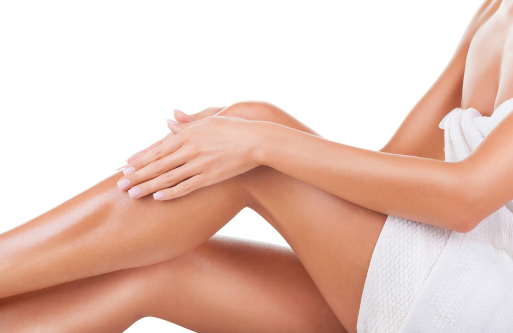
Little, rough bumps can appear on the upper arms, thighs, cheeks, and buttocks due to a skin disorder called keratosis pilaris. Despite its nickname, “chicken skin,” keratosis pilaris is not contagious and cannot be passed from person to person.
Keratosis pilaris is a hereditary condition that can be handed down through families. A wide range of ages, from toddlers to senior citizens, can be affected, and the effects can be long-lasting. There is currently no cure for keratosis pilaris, however the condition can be managed effectively. Keratosis pilaris laser hair removal using an IPL laser is one option.
The skin on your body can sometimes look like sandpaper, and if you have an itchy rash on your arms, legs, back, or chest, you might have Keratosis Pilaris (KP). This is a common but little-known skin condition that causes hard bumps with a red appearance. It’s common in toddlers but can affect people at any age. Read on to learn more on red light therapy for keratosis pilaris/ipl keratosis pilaris reddit.

Ipl Laser For Keratosis Pilaris
Igot to the year 2015 in my phone’s camera roll when I decided to finally call it quits. At first, it was kind of shocking to me that I couldn’t find a true “before” shot of my keratosis pilaris—the small, red bumps on my arms have been a source of insecurity since middle school. But as I continued to scroll through my archives in the name of journalistic duty, my initial frustration began to give way to understanding: From my clothing choices to subtle camera angles (and the occasional Facetune), I’ve just gotten really good at hiding the evidence of my KP.
And perhaps that’s partly because until rather recently, I had resigned myself to the fact that treating this chronic skin condition was hardly worth the effort. In addition to being incredibly common (up to 40 percent of adults deal with it to varying degrees),1 keratosis pilaris is quite harmless. Most of the products and techniques I’ve tried require a lot of diligence for disappointingly modest results. Since it’s much more of an aesthetic nuisance than a physical one, covering it up, even if semi-subconsciously, has always been much more convenient than slathering myself with products day and night.
For me, any worthwhile treatment would have to be quick and permanent, or close to it.
Fast-forward to earlier this fall, when, while undergoing a laser hair removal treatment, I had an epiphany. Keratosis pilaris is a genetic condition that affects the hair follicle: It’s characterized by the skin producing too much keratin, which then blocks the hair follicle, resulting in those signature bumps. In other words, it’s kind of like having a bunch of tiny ingrown hairs all the time—so couldn’t laser hair removal theoretically correct the issue?
My hunch, it would turn out, wasn’t totally off-base—at least according to some light Googling and Will Kirby, MD, the Chief Medical Officer at LaserAway. “[With keratosis pilaris], hair follicles are retained in the skin and cause mild inflammation,” he explains. “As such, in some cases, patients that suffer from KP can see improvements in the condition after getting laser hair removal treatments.”
There’s the caveat: While it might help, there’s no guarantee that laser hair removal can significantly improve that bumpy skin texture. But that wasn’t going to stop me from at least giving it a try.
Read on to see how laser hair removal helped with my KP.
How It Works
A little laser hair removal 101: The laser targets melanin in the hair follicle, converting into heat that damages the follicle and prevents future hair growth. Since keratosis pilaris affects the skin around the hair follicle, laser hair removal isn’t exactly a home run treatment for eliminating KP entirely. “KP can be genetic but it can also be associated with pregnancy, diabetes, and atopic dermatitis, so sometimes the best treatment for KP is to treat the underlying condition,” says Kirby.
But since KP can cause a lot of ingrown hairs—which make the bumps look worse—it stands to reason that eliminating that hair growth would at least help smooth things out a little, right?
Pros and Cons of Laser Hair Removal for KP
As stated above, laser hair removal is thought to help KP because it stops hair growth, therefore, decreasing the coiled ingrown hairs that cause the bumps. Many people have great results and will see fast results that last. It is important to note, however, no studies are showing that laser hair removal can cure KP, so it may not work for everyone.2
Even though laser hair removal is safe for most, the Mayo Clinic says there are some risks depending on a number of factors— including your skin color, hair color, and skin type.3 Some people may experience skin irritation including redness and swelling. Other side effects include pigment changes or excessive hair growth, especially for people with darker skin tones. Talk to your doctor to make sure laser hair removal is a good fit for you.
It is also important to note that laser hair removal requires multiple treatments and, depending on the size of the area treated, and it can get quite expensive.
The Results
:max_bytes(150000):strip_icc():format(webp)/slide-2-b613554360c442488583f46b31a47302.jpg)
After two treatments at LaserAway (which has the double benefit of having top-notch staff and a location very close to my home) my arm hair has been reduced to soft peach fuzz—and my skin, while not perfectly smooth, is certainly less angry-looking. As fate would have it, my smooth-armed technician has also dealt with KP, and reassuringly told me during my first appointment that she saw improvement through laser hair removal almost right away.
Again, I’ve gotten laser hair removal before, so the treatment itself feels relatively routine. My technician marks off my arms with chalk (which makes it easier for her to track each area and also avoid my tattoos, which can be damaged by the laser), and does a single pass over each arm. For those curious about what it feels like, imagine the sensation of a staticky shock or a rubber band snapping on your arm—not particularly painful, but not exactly enjoyable either.
Either way, as I took my first set of progress photos—proudly flashing my arms for the camera for the first time in (apparently) a long while—I was pleasantly surprised when my first instinct wasn’t to open Facetune, but Instagram. I toggled right past the filters and tapped “share,” remaining bumps and all.
Red Light Therapy For Keratosis Pilaris
Light sources such as sunlight, red light, blue light devices activate a chemical in the photosensitizer that temporarily treats keratosis. This type of photodynamic therapy can be useful for severe cases when you need relief quickly
If you’ve contracted a skin condition that seems like acne or small pimples but doesn’t respond to the usual treatments, you may very well have keratosis pilaris. A visit to the dermatology office can “clear things up” about the nature of your skin condition and how to treat it. Keratosis pilaris is rough bumps on your skin that some say look like the skin of a plucked chicken. If you do have keratosis pilaris, don’t be alarmed. It’s a very common skin condition made up of clumps of dead skin cells on the surface of your skin. These clumps or plugs usually appear on your upper arms and the front of your thighs. Children may develop this skin condition on their faces and cheeks.
Symptoms of Keratosis Pilaris
Your arms, thighs, and other areas where you develop keratosis pilaris may become dry and itchy. If your skin is dry, the effects of keratosis are more noticeable. People who live in a dry climate find they only develop this skin condition in the winter when it’s drier. If you live in a warm, dry climate and swim throughout the year, you may experience this condition year-round. If you spend a lot of time in direct sunlight, you may have a solar keratosis variation.
This condition, also known as actinic keratosis, appears as scaly, rough patches on your face and head. Older people are more at risk of experiencing this skin condition, which is harmless. However, there is a small risk of becoming skin cancer, so you should visit your dermatologist to receive a full examination and any recommended treatment.
Treating Keratosis Pilaris
There is no known cure for keratosis pilaris. But don’t lose heart if you have it. In some cases, it goes away on its own. In others, its symptoms can be controlled and treated. Talk to your dermatologist about the treatments that may work best for you. Fortunately, there are many skincare recipes and treatment options available to you.
People get the condition for a variety of reasons. Plus, different people have different types of complexions. These and other factors prohibit a one-size-fits-all treatment. No therapy for keratosis pilaris is effective for everyone across the board. So you may have to try several treatments before you find some that work for you.
One general treatment to prevent dry skin is using a mild cleanser that doesn’t contain soap, such as Cetaphil or Dove. Exfoliation is also helpful for getting rid of the small plugs of keratin in the top layers of your skin. The best results are typically found by combining therapies.
You can see improvement by adhering to an ongoing skincare plan. Many people respond well to skincare programs designed to treat keratosis. Just remember that treatment has to be ongoing. If you don’t use the lotions or therapeutic creams as directed, the condition can return. In some cases, a complete clearing of keratosis isn’t possible even when diligently following the treatment plan.
Prescription Medication for Keratosis
Dermatology doctors may prescribe a topical steroid cream like Cloderm or Locoid Lipocream. These are typically prescribed for seven to 10 days, applied one to two times daily to inflamed or red areas. Once the swelling is reduced successfully, you can treat the remaining rough, dry bumps with a mixture of urea cream and salicylic acid. Another effective treatment for keratosis is the intermittent application of topical retinoids. These are stronger medicines that are applied weekly or biweekly. When your condition starts to clear, milder creams may be used as part of an ongoing regimen.
Co-Occurring Conditions
If you are experiencing hyperpigmentation, which is ongoing skin discoloration, you can treat it with a fading cream that contains hydroquinone (HQ), azelaic acid, and kojic acid. If your skin discoloration is more extreme, consult your dermatologist for specific treatments. You may need a stronger concentration of HQ — possibly six, eight, or 10 percent. Be aware that a higher concentration of HQ can cause irritation and possibly even ochronosis, during which your skin may turn a bluish-black color. Topical immunomodulators are also effective at treating keratosis. These include creams with the active ingredient tacrolimus or pimecrolimus, which go by Protopic and Elidel’s brand names. These treatments are typically used if you have severe inflammation or redness in the affected area.
Light Therapy for Keratosis
Dermatologists may also suggest a two-step combination of a light source and topical photosensitizer for temporary treatment of keratosis. Light sources such as sunlight, red light, blue light devices activate a chemical in the photosensitizer that temporarily treats keratosis. This type of photodynamic therapy can be useful for severe cases when you need relief quickly. Unfortunately, it doesn’t offer long-term treatment, but light therapy can be used in conjunction with other, more lasting treatments.
Surgical Care for Keratosis Pilaris
If creams and other treatments aren’t effective in treating your keratosis, your dermatologist may recommend surgical treatments, such as a minor surgery called gentle acne extraction. This surgery is performed with a diabetic lancet to cut the skin. Then instruments are used to remove the trapped coiled hairs or keratin plugs formed below the skin, causing your keratosis. Your dermatologist can perform several surgical procedures as well as more conventional treatments right in the office. These include:
- Dermabrasion;
- Microdermabrasion;
- Abrasion, which uses a synthetic diamond to exfoliate your skin and a vacuum to remove dead skin cells;
- Chemical peels;
- Photodynamic therapy;
- Laser treatments;
- Blue light therapy;
- Pulsed light instruments;
- Surgical extraction of trapped hairs or keratin plugs.
There are possible side effects related to surgery and the other treatments listed above for keratosis. The risks include skin tautness, redness, swelling, skin sensitivity, and slight bruising. All these side effects are temporary and relatively minor.
Preventing Keratosis Pilaris
Your dermatology specialist may provide you with in-home treatments to keep your keratosis from returning. Proceed with care and make sure you follow the dermatologist’s directions and maintain the prescribed therapy regimen. At-home therapies include:
- Retinol creams;
- Exfoliation pads such as Buf-Puff;
- Topical emollients;
- Gentle suction exfoliation, such as Vacubrasion;
- Glycolic acid peels with a strength of 10 to 20 percent.
You can prevent your keratosis from becoming worse and possibly prevent it entirely by taking measures to prevent dry skin. Some of these measures include frequently applying emollients and cleaning with mild soaps and cleansers that don’t contain soap.“”★ ★ ★ ★ ★Highly recommend visiting and get checked out. The process was smooth, and the doctor has a lot of knowledge on the subject.
Keratosis Pilaris FAQ
What Is Keratosis Pilaris?
Keratosis pilaris (KP) is commonly referred to as a harmless skin condition that leads to dry, rough patches and tiny bumps. They typically appear on the thighs, upper arms, cheeks, or buttocks. These bumps do not tend to itch or hurt. This skin disorder cannot be cured or prevented. However, it can be managed with moisturizers and prescription creams to improve the appearance of the skin. KP normally disappears by the age of 30.
What Are the Causes of Keratosis Pilaris?
This skin disorder is believed to result from the buildup of keratin, which is a hard protein that protects the skin from harmful infections and harmful substances. Keratin forms a scaly plug that blocks the opening of the hair follicle. Normally, many plugs form, causing patches of bumpy and rough skin. It is still to be discovered why keratin builds up. Although, it may occur because of genetic diseases or in association with other skin conditions. Keep in mind that dry skin has been shown to worsen keratosis pilaris symptoms.
How to Get Rid of Keratosis Pilaris?
Keratosis pilaris, also known as chicken skin, cannot be cured, but you can still manage its symptoms if you do not feel confident about your skin looks. This skin condition typically disappears by itself as a person ages. KP skin condition is generally treatment-resistant, but several methods can ease its symptoms. It is also possible to address this skin disorder in-home settings using a warm bath, exfoliation, coconut oil, and humidifiers. People who got exposed to KP should also avoid wearing tight clothes.

Importance Keratosis pilaris (KP) is a common skin disorder of follicular prominence and erythema that typically affects the proximal extremities, can be disfiguring, and is often resistant to treatment. Shorter-wavelength vascular lasers have been used to reduce the associated erythema but not the textural irregularity.
Objective To determine whether the longer-wavelength 810-nm diode laser may be effective for treatment of KP, particularly the associated skin roughness/bumpiness and textural irregularity.
Design, Setting, and Participants We performed a split-body, rater-blinded, parallel-group, balanced (1:1), placebo-controlled randomized clinical trial at a dermatology outpatient practice of an urban academic medical center from March 1 to October 1, 2011. We included all patients diagnosed as having KP on both arms and Fitzpatrick skin types I through III. Of the 26 patients who underwent screening, 23 met our enrollment criteria. Of these, 18 patients completed the study, 3 were lost to or unavailable for follow-up, and 2 withdrew owing to inflammatory hyperpigmentation after the laser treatment.
Interventions Patients were randomized to receive laser treatment on the right or left arm. Each patient received treatment with the 810-nm pulsed diode laser to the arm randomized to be the treatment site. Treatments were repeated twice, for a total of 3 treatment visits spaced 4 to 5 weeks apart.
Main Outcomes and Measures The primary outcome measure was the difference in disease severity score, including redness and roughness/bumpiness, with each graded on a scale of 0 (least severe) to 3 (most severe), between the treated and control sites. Two blinded dermatologists rated the sites at 12 weeks after the initial visit.
Results At follow-up, the median redness score reported by the 2 blinded raters for the treatment and control sides was 2.0 (interquartile range [IQR], 1-2; P = .11). The median roughness/bumpiness score was 1.0 (IQR, 1-2) for the treatment sides and 2.0 (IQR, 1-2) for the control sides, a difference of 1 (P = .004). The median overall score combining erythema and roughness/bumpiness was 3.0 (IQR, 2-4) for the treatment sides and 4.0 (IQR, 3-5) for the control sides, a difference of 1 (P = .005).
Conclusions and Relevance Three treatments with the 810-nm diode laser may induce significant improvements in skin texture and roughness/bumpiness in KP patients with Fitzpatrick skin types I through III, but baseline erythema is not improved. Complete treatment of erythema and texture in KP may require diode laser treatment combined with other laser or medical modalities that address redness.
Trial Registration clinicaltrials.gov Identifier: NCT01281644
Keratosis pilaris (KP) is a common hereditary, benign disorder of unknown etiology1 that is frequently seen in conjunction with atopy. The hereditary pattern of this skin disorder is thought to be autosomal dominant without a known predisposition based on race or sex.2 Keratinaceous plugging of follicles results in markedly visible papules, often involving the lateral and extensor aspects of the proximal extremities but sometimes also the face, buttocks, and trunk.3 Perifollicular erythema is routinely notable.4 Topical treatments for KP include emollients, exfoliants, and anti-inflammatory agents, such as urea, salicylic acid, lactic acid, topical corticosteroids, topical retinoids, and cholecalciferol. Because most patients obtain limited benefit from these treatments, less conventional treatments, including phototherapy and lasers, have been explored. Among lasers, the 532-, 585-, and 595-nm vascular devices have been used with modest success, particularly in reducing redness.5–8 Longer-wavelength lasers have not been studied for the treatment of KP, and lasers have not been shown to be successful for treating the textural components of KP. Our study investigates the effectiveness of the longer-wavelength 810-nm diode laser for color and texture of upper extremity KP.
We performed a split-body, parallel-group, placebo-controlled randomized clinical trial with an allocation ratio of 1:1 and a block size of 2 at an urban academic medical center. The unit of randomization was the individual unilateral upper extremity. The study was approved by the institutional review board of Northwestern University. All participants provided written informed consent.
Patients were recruited from a dermatology practice at Feinberg School of Medicine, Northwestern University, and the surrounding community. Inclusion criteria consisted of age 18 to 65 years, good health, Fitzpatrick skin types I to III, and a diagnosis of KP on both upper extremities. We excluded patients who had received any laser therapy to the arms in the 12 months before recruitment, with a concurrent diagnosis of another skin condition or malignant neoplasm, with a tan or sunburn over the upper arms in the month before recruitment, with open ulcers or infections at any skin site, or who were using topical or oral photosensitizing medications.
When potential participants called or e-mailed the clinic for possible inclusion in the study, they underwent prescreening (performed by O.I.) over the telephone using the aforementioned inclusion and exclusion criteria. Once enrollment criteria were met, patients were scheduled for a total of 4 visits, 4 to 5 weeks apart, in the Department of Dermatology, Feinberg School of Medicine.
On the patient’s first visit, one of us (O.I.) reviewed the inclusion and exclusion criteria. After the patients provided written informed consent, they separately rated redness and roughness/bumpiness on each arm using a scale of 0 (least severe) to 3 (most severe) for a total maximum score of 6 per patient per arm. Next, patients were randomized into 2 groups as described below, and baseline standardized digital photographs were obtained. Each patient received treatment using the 810-nm pulsed diode laser to the arm randomized to be the treatment site. After laser treatment, both sides were treated with topical petrolatum. Treatments were repeated twice for a total of 3 treatment visits, with visits spaced 4 to 5 weeks apart. At the fourth and final visit, 12 to 15 weeks after the initial visit, the patients again rated disease severity as previously described. At this last visit, 2 blinded dermatologists (S.Y. and M.A.) also rated the roughness/bumpiness and redness of the treatment and control arms separately using the same scales, and digital photographs were again obtained.
Patient screening and enrollment were performed by one of us (M.D.), as were random sequence generation and concealment (R.K.), which were conducted by coin toss of the same fair coin, with the outcomes (1 or 2) recorded separately on individual paper cards then placed in sealed, opaque, consecutively numbered envelopes. Each patient was assigned to one of 2 groups (by W.D.). Patients in group 1 were designated to receive laser therapy on the right arm, and those in group 2 were assigned to receive laser therapy on the left arm. All study treatments were delivered by the same clinician (D.B.).
All study treatments used the 810-nm pulsed diode laser. A lidocaine and prilocaine–based cream was applied to the arms 30 to 60 minutes before treatment and washed off before treatment. Laser therapy was performed on the treatment side at a fluence of 45 to 60 J/cm2 (to convert to gray, multiply by 1) (depending on Fitzpatrick skin type) and a pulse duration of 30 to 100 milliseconds, with precise settings selected to be just below the patient’s threshold for purpura. Each treatment session entailed 2 nonoverlapping passes separated by a 1-minute delay. The patient was then instructed to minimize sun exposure and apply sunscreen with a sun protection factor of 50 to the treatment area daily until the next visit.
The primary outcome measure was the difference in disease severity score, including redness and roughness/bumpiness, between the treated site and the control site as rated by the blinded dermatologists at 12 weeks after the initial visit. This scale was not validated because no relevant validated scale was available. However, raters were trained on the use of the study scale, and before the review of study images, they were asked to rate archival skin images on the same 4-point qualitative subscales used in the study. Raters reviewed and rated archival images separately and then reconciled their ratings through face-to-face forced agreement, with the process repeated until concordance was achieved between raters and their separately rated scores were consistently equivalent.
During the evaluation of study data, forced agreement was used to reconcile blinded ratings. The secondary outcome measure was the change from baseline in disease severity of each arm as rated by the patients.
Power Analysis and Sample Size
Assuming an SD of change of 0.84, a sample of 20 patients had 80% power to detect median differences (or median changes) in severity scores of 0.5. We assumed a 2-sided test and type I error rate of 5%.
We used the Wilcoxon signed rank test to compare the magnitude of change from baseline between treatment and control for all patient ratings (redness, roughness/bumpiness, and overall score). Blinded dermatologists’ ratings of the treatment and control sides were also compared using the Wilcoxon signed rank test.
Patient Baseline Demographic Characteristics
The study was conducted during a 7-month period from March 1 to October 1, 2011. A total of 26 patients underwent screening for our study, and 23 of those patients (46 arms) met our criteria and were enrolled in the study. Of these 23 patients, 18 (36 arms) completed the study and underwent analysis, 3 were lost to or unavailable for follow-up, and 2 voluntarily withdrew owing to inflammatory hyperpigmentation after the laser treatment. The demographic characteristics of our patients are presented in the Table. At baseline, patients rated the severity of the roughness/bumpiness in the texture of their arm test sites at a median score of 1.5 (interquarile range [IQR], 1-2) and the severity of the erythema of their arm test sites at a median score of 2.0 (IQR, 1-2). (The maximum score for both ratings was 3.0.)
At follow-up, the median redness score assigned by the blinded raters for the treatment and control sides was 2.0 (IQR, 1-2), a null difference (Figure 1). The median roughness/bumpiness score was 1.0 (IQR, 1-2) for the treatment sides and 2.0 (IQR, 1-2) for the control sides, a difference of 1 (P = .004) (Figure 1). The median overall score combining erythema and roughness/bumpiness was 3.0 (IQR, 2-4) for the treatment sides and 4.0 (IQR, 3-5) for the control sides, a difference of 1 (P = .005) (Figure 1).
Patient Self-assessment Scores
At follow-up, patients’ self-reported median erythema rating for the control sides did not change from the baseline score of 2.0 (IQR, 1-2), but the self-reported median erythema score for the treatment side decreased from 2.0 to 1.5 (IQR, 1-2), a nominal difference that was not statistically significant (P = .13) (Figure 2). The median roughness/bumpiness score for the control sides increased from 1.5 to 2.0 (IQR, 1-2) and for the treatment sides decreased from 1.5 to 1.0 (IQR, 1-2). The 1-point decrease in roughness/bumpiness in the treatment arm compared with the control arm was significant (P = .008) (Figure 2). The overall score (erythema and roughness/bumpiness) for the control sides increased from 3.5 to 4.0 (IQR, 3-4), and for the treatment arm decreased from 3.5 to 2.5 (IQR, 2-4), with the cumulative difference of 1.5 points being significant (P = .005) (Figure 2).
We found no unexpected adverse events associated with laser treatment. Two participants developed inflammatory hyperpigmentation after laser treatment and chose to withdraw from the study. These patients were instructed to continue sun-protective measures to their affected extremities, and in both cases hyperpigmentation completely resolved within 3 months.
We investigated the effectiveness of the 810-nm diode laser in the treatment of KP. After 3 treatments spaced 4 to 5 weeks apart, blinded dermatologist ratings and patient self-report indicated significant improvements in skin texture and roughness/bumpiness when compared with baseline However, neither raters nor patients detected a significant change in erythema.
Most topical treatments for KP, including emollients, corticosteroids, and retinoids, are of limited effectiveness.9 Light-based treatments have typically entailed use of vascular lasers, like the application of a 532-nm potassium titanyl phosphate laser to treat a case of resistant facial KP by Dawn et al.5 Repeated treatments resulted in a marked improvement in erythema and some clearance of papules. A study of 12 patients using the 585-nm pulsed-dye laser6 found improvement in erythema but not in roughness/bumpiness. A similar report7 described a case in which multiple treatments with a 595-nm pulsed-dye laser induced marked improvements in facial erythema, patient satisfaction, and quality of life. A study of 10 patients treated with a 595-nm pulsed-dye laser8 confirmed these results.
To our knowledge, our study is the first of its kind to investigate the use of a longer-wavelength laser, the diode laser, in the treatment of KP. More important, our results are the first from a clinical trial that demonstrate the effectiveness of laser treatment of the textural abnormality and roughness/bumpiness associated with KP. The data from our investigation suggest that the 810-nm diode laser is a particularly promising and effective treatment for the nonerythematous variants of KP. The variant of KP known as keratosis pilaris alba, which presents mostly as follicular papules, may be highly responsive to this laser modality.10 The variant that includes perifollicular erythema with follicular papules, keratosis pilaris rubra,9,10 may best respond to joint treatment with diode and vascular lasers, with the former improving texture and the latter addressing erythema.
We have theoretical reasons for selecting the 810-nm diode laser and the settings used in this study. Specifically, KP is an inflammatory condition of vellus hair follicles. Compared with terminal hair, vellus hair is relatively deficient in melanin (ie, has less chromophore) and smaller in diameter (ie, has shorter thermal relaxation time). Based on the theory of selective photothermolysis, these features would be consistent with a thermal relaxation time of approximately 50 milliseconds, which means that a pulse duration of less than 50 milliseconds, such as the 30 milliseconds used in this study, would be appropriate for treatment. Because of a substantial lack of chromophore, the fluence required for photothermal destruction of a vellus hair follicle is 40 to 45 J/cm2, greater than that for a terminal hair. Ideally a highly absorbing wavelength such as 695 nm would be the best to treat vellus follicles, but this wavelength is absorbed by epidermal pigment in darker skinned individuals before it can reach deeper targets, such as the stem cells in the bulge region of the follicles. Similarly, 1064 nm is not highly selective for melanin, and we know that the vellus follicle has little melanin to begin with. As a consequence, the 810-nm wavelength appears to be the best choice because its depth of penetration is sufficient, it has selectivity for melanin, and it is compatible with a pulse duration of 30 milliseconds.
In terms of adverse events, our study found that treatment with the 810-nm diode laser was safe and not associated with any serious or unexpected adverse events. Although 2 patients (9%) developed bothersome inflammatory hyperpigmentation after laser treatment, resulting in their withdrawal from the study, these sequelae resolved completely in the medium term. Further counseling about the need for sun protection and avoidance of tanning during the period of laser treatment may mitigate the risk for posttreatment inflammatory hyperpigmentation in the future.
A limitation of our study is that enrollment was restricted to participants with Fitzpatrick skin types I to III. The exclusion of darker skin types was not incidental but rather designed to minimize the risk for posttreatment inflammatory hyperpigmentation, which is more common after laser procedures in patients with Fitzpatrick skin types IV to VI. That posttreatment inflammatory hyperpigmentation was observed in this study despite careful patient selection suggests that this precaution was appropriate. Regardless, patients with darker skin types can indeed be treated safely with the diode laser if gentle settings are used. Once this treatment paradigm is optimized, such broader application will likely be appropriate and feasible. One protective benefit of the current treatment settings was that they were deliberately below the threshold for purpura and thus designed to avoid bruising, which can resolve with tan pigmentation, particularly in darker skin. To the extent that the 810-nm diode laser has hair-removing activity, this treatment may be inappropriate for patients who do not want hair loss at the site of their KP. Finally, although incidental reports from some participants previously in this study have indicated that they have maintained textural benefits for more than a year, it remains to be seen to what extent these improvements are maintained over the longer term. To the extent that laser treatment may significantly modify hair growth in abnormal vellus hair follicles initially induced by genetic predisposition, improvement may be long lasting. This result would then be parallel to the case of traditional hair removal, in which posttreatment long-term remission of coarse terminal hairs and the corresponding pseudofolliculitis is often observed.
However, this study was not designed to assess long-term improvement, and additional studies would need to be performed to systematically measure the duration and likelihood of persistent benefits. The present study only provides proof of concept and indicates that improvement of the textural abnormalities associated with KP is possible after treatment with an 810-nm diode laser.
By objective and subjective measures, we found that, among lighter-skinned persons, serial treatment with a long-pulsed 810-nm diode laser at subpurpuric levels provided medium-term improvement in KP, particularly for the associated roughness/bumpiness and textural irregularity. Combined with preexisting data about the utility of vascular lasers for the reduction of KP-associated erythema, this finding suggests that laser treatment may comprehensively address the clinical manifestations of KP in selected patients. Future studies may assess the durability of these responses and the comparative effectiveness of different long-wavelength lasers.
Ipl Keratosis Pilaris Reddit
Ipl Keratosis Pilaris (KP) is a genetic skin condition that presents as tiny, red bumps on the upper arms and thighs. It can be itchy, but it’s not contagious or painful.
The bumps are caused by keratin — a protein that protects the skin from moisture loss — getting trapped in hair follicles. It’s not clear what causes KP, but it can run in families and occur with other conditions such as eczema and psoriasis.
One of the most common methods for treating KP is with a chemical peel. A dermatologist will apply a chemical solution to your skin that causes it to blister and peel off within three days. The results are usually quite dramatic and permanent
ipl keratosis pilaris reddit
The first thing that comes to mind when you think of a skin condition is probably the kind of breakouts, rashes, and other conditions you get on your face. But there are many other types of skin conditions that can affect any part of your body, including your scalp.
One such condition is ichthyosis vulgaris, more commonly known as fish scale disease or ichthyosis. This is a genetic disorder that affects approximately 1 in every 250 people worldwide and causes a dry, scaly skin rash to form over large areas of the body.
The rash most commonly appears on the upper arms, back, chest and thighs but can also appear on the face, neck and hands. It usually appears at birth or within the first few months and typically worsens during puberty before improving again later in life.
Ipl keratosis pilaris reddit. Ipl is a skin condition that causes hard bumps on the back of the arms and legs, and sometimes on the face. It’s more common in people with dry skin, but it can occur in anyone. The bumps are often mistaken for goosebumps or pimples.
What is ipl?
Ipl stands for ichthyosis-pilaris lichen planus, which is a rare genetic skin disorder that causes small, painful bumps on the upper arms and thighs. It also affects the cheeks, forearms, knees and buttocks.
The bumps are usually pale pink to white and look like goosebumps when touched. They can be as small as a pinhead or as large as 2 millimetres (0.08 inch) across. Each bump contains a core of hair follicles (tiny structures that secrete oils). Some people with ipl may also get redness of their skin after shaving or exposure to sunlight (photosensitivity).
How common is it?
ipl affects around one in every 1,000 people worldwide but its exact prevalence isn’t known because most cases aren’t reported to doctors or recorded in medical journals.
The skin is the largest organ of the body, so it’s no surprise that skin imperfections, blemishes, marks and lesions can happen. Many skin conditions not harmful to your health, but can be a nuisance, unsightly or even embarrassing. Keratosis Pilaris is one such condition, and treating Keratosis Pilaris is simple.
It’s very common and completely harmless but if you suffer with it, can be something of a less than welcome part of your life, due to its sometimes, unattractive appearance. If you have small pimples on the skin that look like permanent goose bumps on areas of the body such as the back of your arms, legs, bottom and even the back, face, eyebrows and scalp, which sometimes get itchy or red, you may have Keratosis Pilaris.
The condition occurs when there is a build-up of a substance called Keratin, a natural protein, which in fact is the main component of the hair as well as healthy skin. This, excess Keratin blocks the openings of the hair follicles, which can cause the small red or white bumps to appear. Keratosis Pilaris also takes the name of “chicken skin” as the skin takes on this appearance. So, no wonder that many people who experience it would like to reduce oreven, eradicate the symptoms.
So, how are we treating Keratosis Pilaris?
Fortunately, at Skin Perfection London, we offer a choice of non-surgical solutions for treating Keratosis Pilaris, painlessly, safely and effectively, from the comfort of our clinic, which is based in the heart of London, between Oxford Street, Harley Street and Bond Street. Treatments can be used alone or combined, for a holistic approach to reducing the chicken skin appearance.
Laser hair removal is a superb way of treating Keratosis Pilaris at its cause. It’s safe, virtually painless and can be permanent! It works by emitting short pulses of light in to the hair follicle, causing it to stop growing hair and to close. This means that it can no longer be blocked by the Keratin and the condition can be drastically improved. The treatment may take up to 9 sessions for optimum results, but can be a long-term solution to this troublesome condition and far better than having to shave, wax or epilate the hair, which can be extremely painful and can exacerbate the symptoms. Laser hair removal is suitable for all skin types and you could see up to 95% permanent reduction in hair growth, so it’s a win-win!
Medical microdermabrasion could be another option. It works by resurfacing the skin and cleaning blocked and congested pores and offers very little downtime or discomfort. At Skin Perfection London, we use the Derma Genesis medical microdermabrasion system, which utilises tiny medical-grade aluminium oxide crystals, which are swept across the skin by a hand-held device. The crystals are then gently sucked back up, bringing with them dirt, debris and dead surface skin cells. This reveals a smoother, clearer and healthier complexion, less prone to becoming congested. Results can be seen after a course of several sessions and your skin expert will explain the treatment programme, along with expected results, at a no obligation consultation, prior to treatment.
Although a harmless condition, Keratosis Pilaris doesn’t have to be endured and at Skin Perfection London, we make it our mission to offer you the most effective, innovative and high-tech device-led treatments to restore smooth, healthy and sexy looking skin, all-year-round.